Pharmaceutical-Grade PDRN · ECM Prime
Cells function properly only in the right environment.
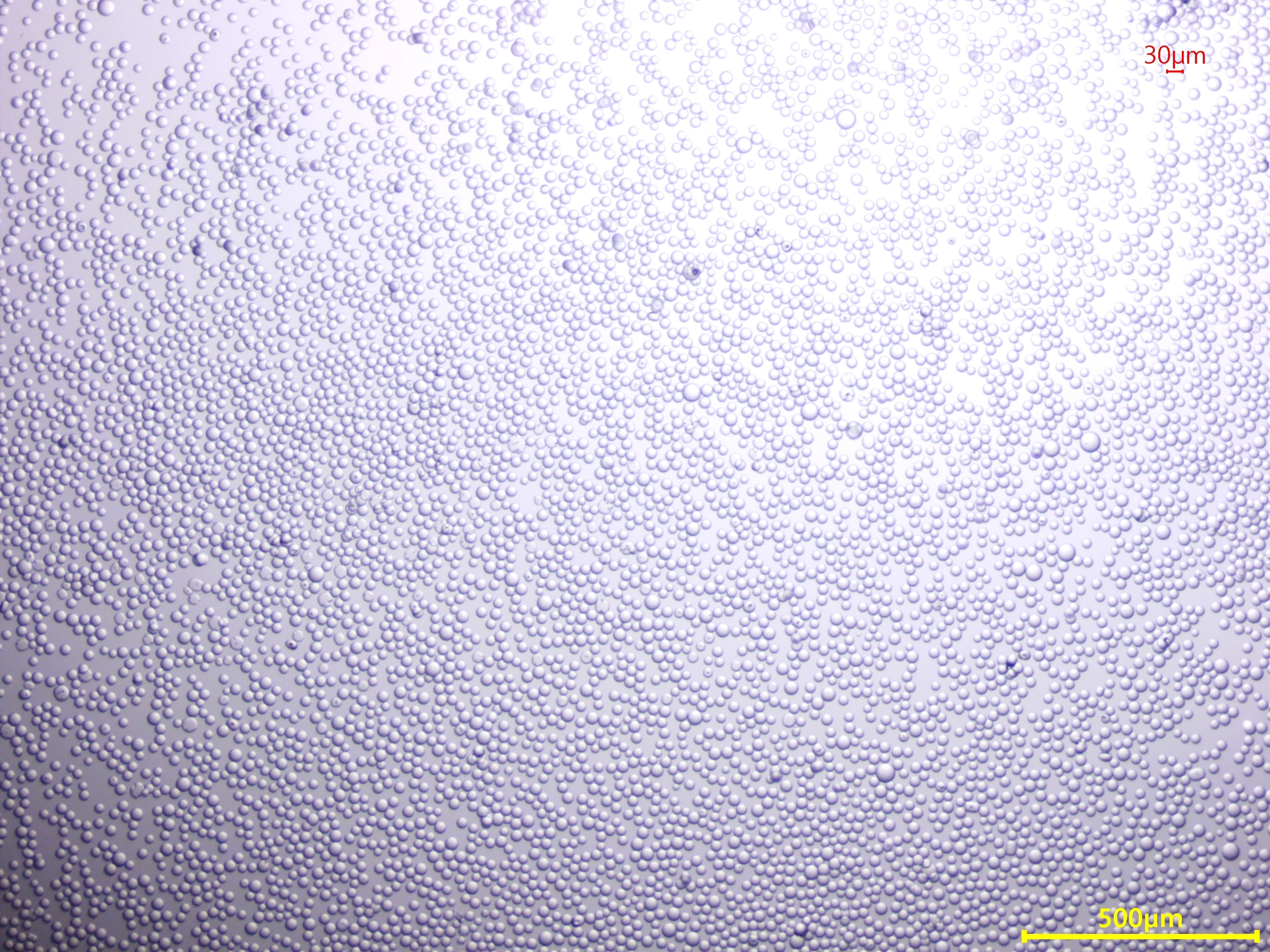
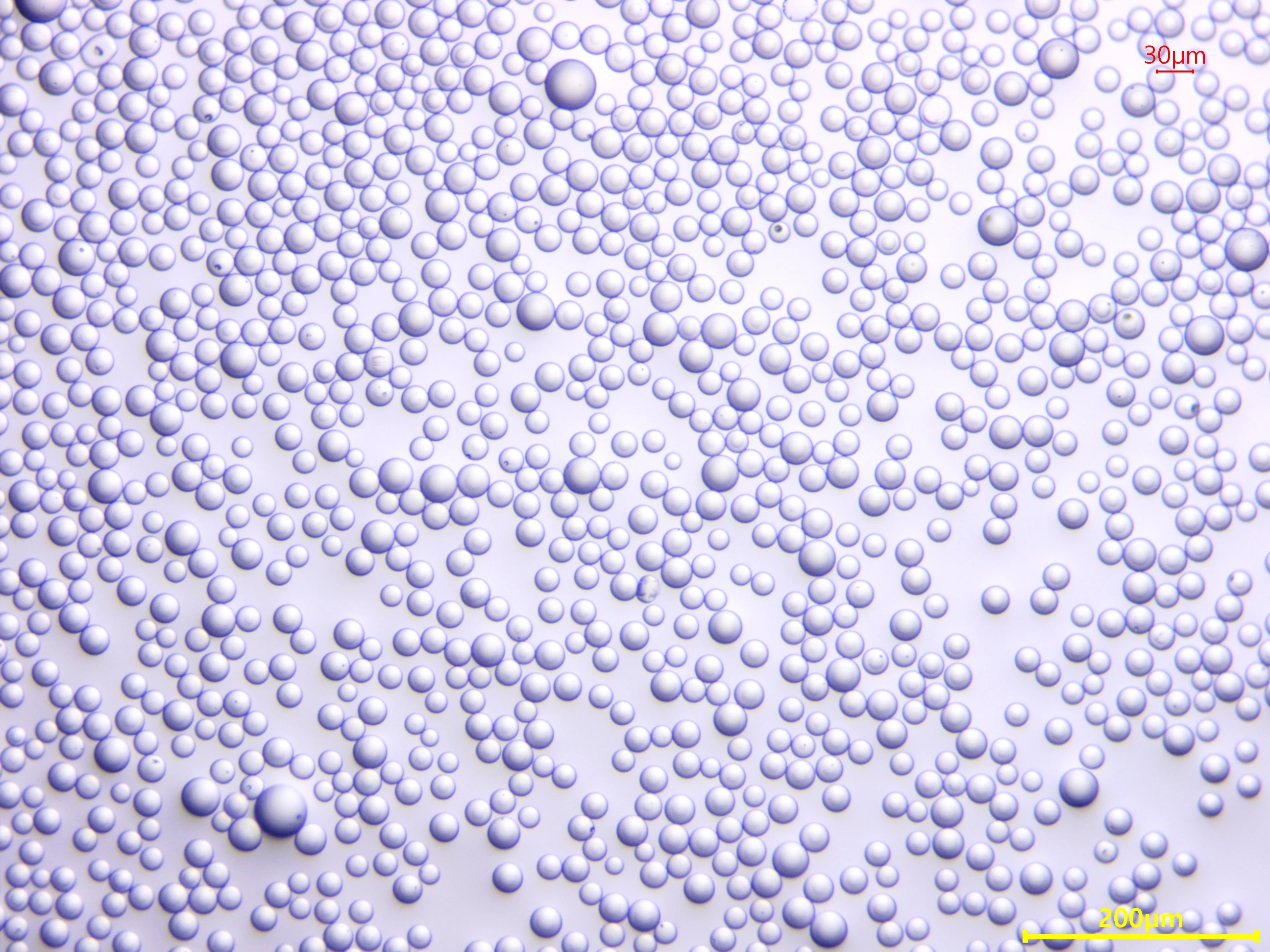
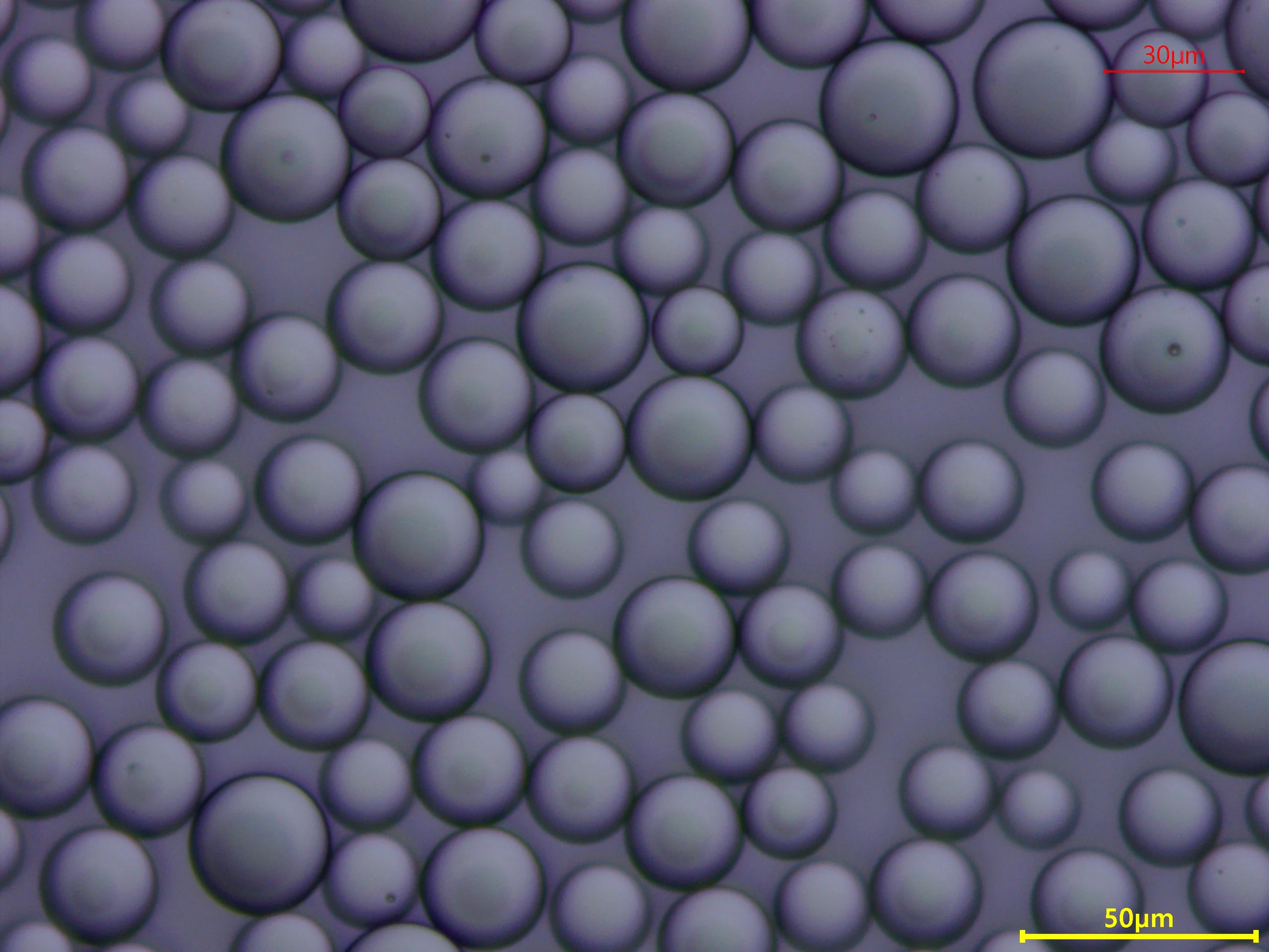
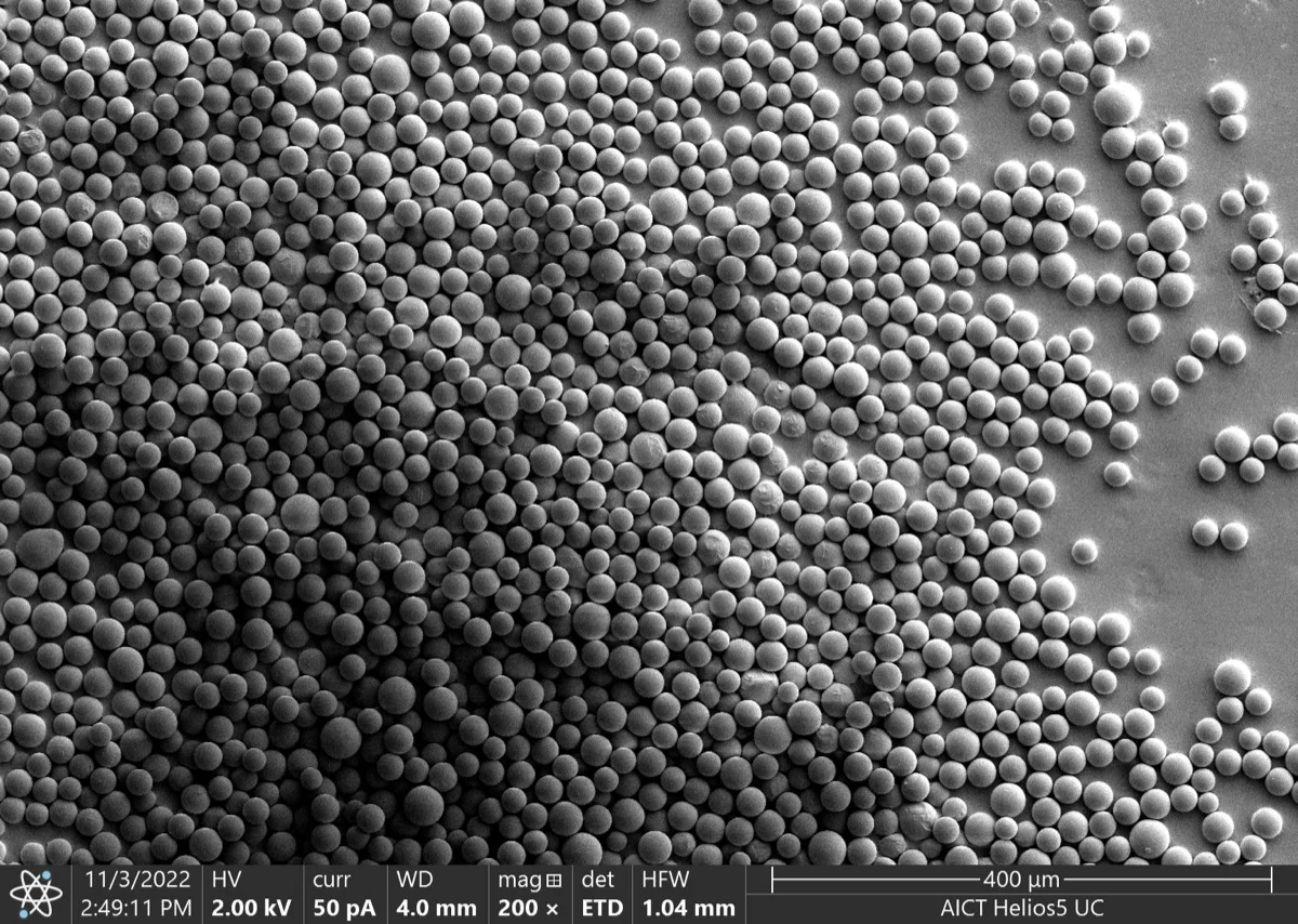
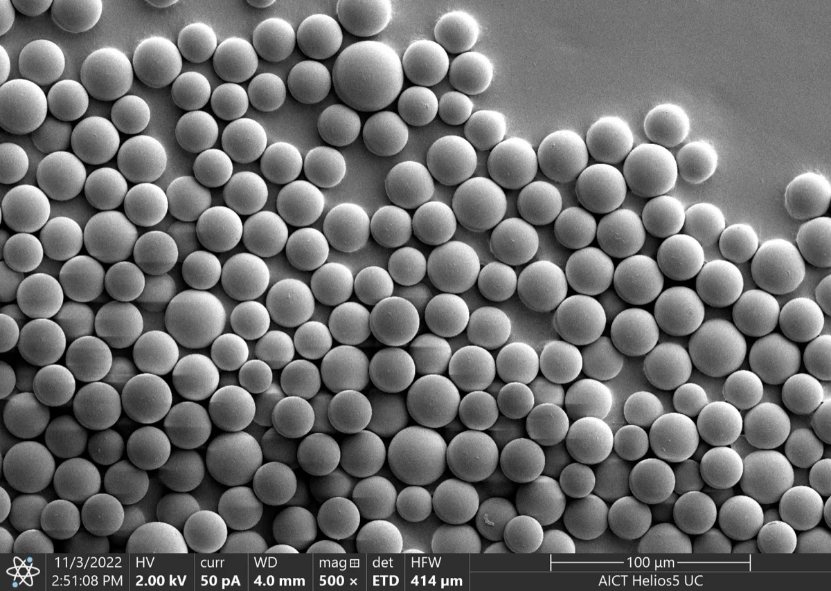
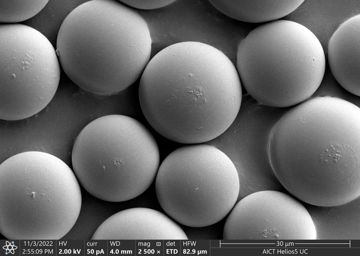
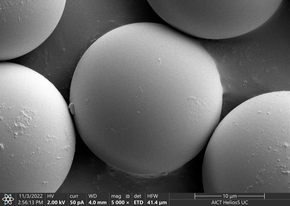
A 4-phase hierarchical design with 50+ components — engineering the entire biological environment for skin conditioning.
01
Foundation
Free Hyaluronic Acid
Establishes the foundational hydration environment of the ECM. Free hyaluronic acid maintains the hydration state of the extracellular matrix and forms the physical foundation where conditioning reactions occur.
02
Activation
PDRN · Amino Acids · B Complex · Signal Peptides
PDRN — supports skin conditioning. 19 Amino Acids — nourish the skin. 9 B Complex + Stable Vit.C — contribute to skin vitality. 9 Signal Peptides — formulated as complex skincare ingredients.
Reference
[3] Colangelo MT et al. Regenerative Medicine, 2020;15(6):1801–1821 · [4] Squadrito F et al. Frontiers in Pharmacology, 2017;8:224
03
Protection
Redox Protection
Glutathione·NAC·Arbutin — block oxidative interference from reactive oxygen species (ROS) generated during skin conditioning. Prevent environmental damage and maintain conditioning quality.
04
Support
Trehalose · Minerals & Electrolytes
Minerals together with trehalose, inositol, and taurine support skin barrier stabilization.
Structure × Environment = Designed Skincare
PDLLA and ECM Prime divide their roles along the time axis, completing a single continuous conditioning pathway.
※ The above references are academic background materials for ingredients and technology, and do not directly substantiate the efficacy of this product.
View Product Details →